SMi Source lesson Atherosclerosis: Heart Failure has the following microlearning topics
1. Role of the Heart and Circulatory System
2. The Heart and Circulation: Anatomy
3. The Heart and Circulation: Physiology
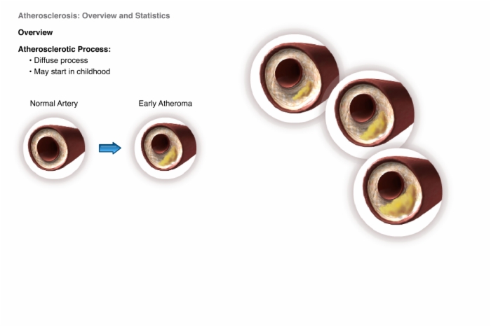
4. Atherosclerosis: Overview and Statistics
5. Atherosclerosis: Risk Factors
6. Atherosclerosis: Diagnostic Tools/Biomarkers

7. Atherosclerosis: Causes
8. Atherosclerosis: Complications
9. Atherosclerosis: Treatments
10. Heart Failure: Overview and Statistics
11. Heart Failure: Risk Factors
12. Heart Failure: Diagnostic Tools/Biomarkers
13. Heart Failure: Causes
14. Heart Failure: Complications
15. Heart Failure: Treatments
Lesson Atherosclerosis: Heart Failure teaches these concepts
Cardiovascular Disease: Atherosclerosis and Heart Failure, Arteries, Veins, Capillaries
Cardiovascular Disease: Atherosclerosis and Heart Failure, Location, Sternum
Cardiovascular Disease: Atherosclerosis and Heart Failure, Appearance, Middle Mediastinum, Pericardium, Parietal Pericardium
Cardiovascular Disease: Atherosclerosis and Heart Failure, Heart is a Double Pump
Cardiovascular Disease: Atherosclerosis and Heart Failure, Cardiac Cycle, Systole, Ventricles, Diastole, Atria
Cardiovascular Disease: Atherosclerosis and Heart Failure, Cardiac Conduction
Cardiovascular Disease: Atherosclerosis and Heart Failure, Cardiac Muscles, Cardiac Myocytes, Heart Muscle Cells
Cardiovascular Disease: Atherosclerosis and Heart Failure, Role of Circulation
Lesson Atherosclerosis: Heart Failure addresses these key points
Heart:
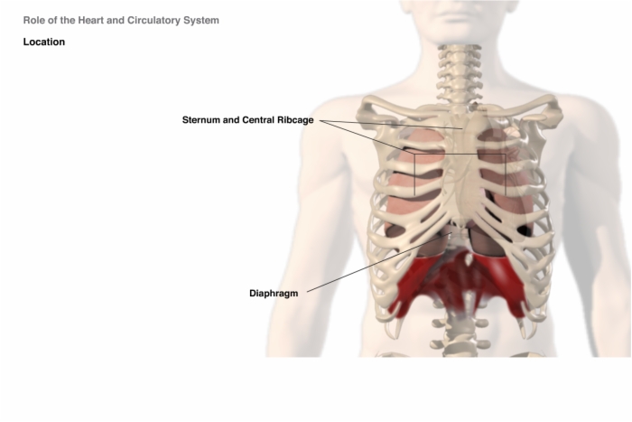
- Located in the middle of the chest between the lungs.
- Along with the great vessels, is protected by the sternum and central part of the thoracic rib cage and rests on the upper surface of the diaphragm.
The apex of the heart is normally located in the fifth intercostal space in the mid-clavicular line.
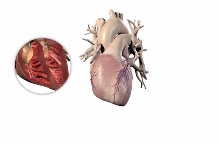
Heart and pericardium occupy a space between the lung cavities, the middle mediastinum.
The fluid between the inner and outer layers of the pericardium allows the inner visceral pericardium to glide smoothly against the outer parietal pericardium.
The heart is a muscular pump that serves two functions:
- The right side collects blood from tissues of the body and pumps it to the lungs which replenish the blood with oxygen.
- The left side collects blood from the lungs and pumps it to the tissues of the body where the oxygen is utilized.
Single cycle of heart's activity can be divided into two basic stages:
- Systole: Contraction and ejection of blood from the ventricles
- Diastole: Ventricular relaxation and filling
Normal Heart Rate: 60-100 beats per minute
Normal Blood Volume: ~ 5 liters (just over 1 ¼ gallons)
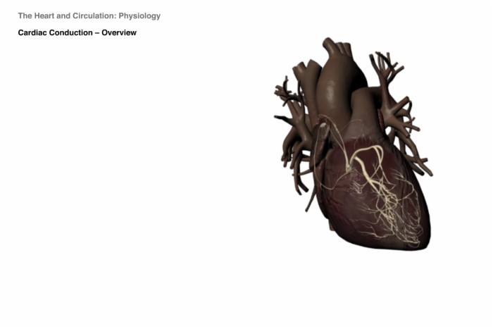
The heart's ability to beat spontaneously, with intrinsic rhythmicity, is made possible through a highly specialized conduction system. Most of these cells have the ability to depolarize spontaneously, which enables them to function as cardiac pacemakers. Electrical impulses begin high in the right atrium in the sino atrial node (the primary pacemaker) and travel through specialized pathways to the ventricles, transmitting the signal to pump.
Cardiac myocytes - one distinguishing feature is the presence of unique proteins in the cell membrane which mechanically connect the cells to one another and also provide a low resistance pathway for the rapid conduction of electrical current which is maintained by ion transport channels.
While cardiac and skeletal muscles are both striated, cardiac cells are smaller, and the action of cardiac cells is spontaneous (involuntary) and intrinsically rhythmic, unlike the action of skeletal muscle which is voluntary.
These rhythmic contractions keep the heart beating in a coordinated and normal rhythm, which in turn keeps blood circulating.
Blood has four general functions:
- Transportation of oxygen, carbon dioxide, nutrients, wastes and hormones
- Regulation of pH, temperature and osmotic pressure
- Protection against foreign substances and pathogens by humoral and cellular immune surveillance
- Prevention of excessive blood loss through the mechanism of clotting
Blood leaves the heart through the major arteries which branch into progressively smaller arteries and arterioles. The smooth muscle cells wrapped around arteries and arterioles play an important role in determining blood pressure. These in turn branch into a network of billions of vessels with the smallest diameter – the capillaries.
Capillaries are only one endothelial cell thick, making them both thin and fragile. The exchange of oxygen, carbon dioxide, and other molecules takes place through the capillary wall.
From the capillaries, blood flows into venules, which then become veins and eventually form the major veins which return blood to the heart.
Lesson Atherosclerosis: Heart Failure is built from these main references. Log into SMi Source for a complete list and details.
Roberts KP, Weinhaus AJ. Anatomy of the Thoracic Wall, Pulmonary Cavities, and Mediastinum. In: Iaizzo PA, ed. Handbook of Cardiac Anatomy, Physiology, and Devices. United States: Humana Press, Inc.; 2005:25-50.
Weinhaus AJ, Roberts KP. Anatomy of the Heart. In: Iaizzo PA, ed. Handbook of Cardiac Anatomy, Physiology, and Devices. United States: Humana Press, Inc.; 2005:51-79.
Mohrman DE, Heller LJ. The Heart Pump. In: Lange Physiology Series. Cardiovascular Physiology. The McGraw-Hill Companies; 2006:47-70.
Laskowski ER. What’s a normal heart rate? MayoClinic.com Website. Sept. 30, 2008. Available at: http://www.mayoclinic.com/health/heart-rate/an01906.
Wilcken DEL. Clinical physiology of the normal heart. In: Oxford Textbook of Medicine, Fourth Edition. Warrell DA, Cox TM, Firth JD, Eds. New York, NY: Oxford University Press, Inc.; 2005;822.
Barnett VA. Cardiac Myocytes. In: Iaizzo PA, ed. Handbook of Cardiac Anatomy, Physiology, and Devices. United States: Humana Press, Inc.; 2005:113-121.
Iaizzo PA. General Features of the Cardiovascular System. In: Iaizzo PA, ed. Handbook of Cardiac Anatomy, Physiology, and Devices. United States: Humana Press, Inc.; 2005:3-11.
Lesson Atherosclerosis: Heart Failure introduces and defines these terms
Pericardium - the heart's covering. A serous membrane containing inner and outler layers with fluid in between.
Cardiac myocytes - heart muscle cells. Cells that are specialized to generate force.